Why Expecting Moms Need to Know the Reasons to Have a Fetal Echocardiogram?
Verified By Dr. Pratima Dash | 05-Sep-2024
Pregnancy is such a magical experience and journey, full of hope and possibilities. But it also brings the responsibility of providing the best care for mother and baby. Fetal echocardiogram is one of the several tests that includes in the list recommended during pregnancy. But this test is for what? What Does It Say about the Well-being of Your Baby? This write-up by Dr. Pratima Dash, Consultant Maternal Fetal Medicine Specialist at Kailash Hospital, Noida addresses the why of a fetal echocardiogram and its importance.
Table of Content
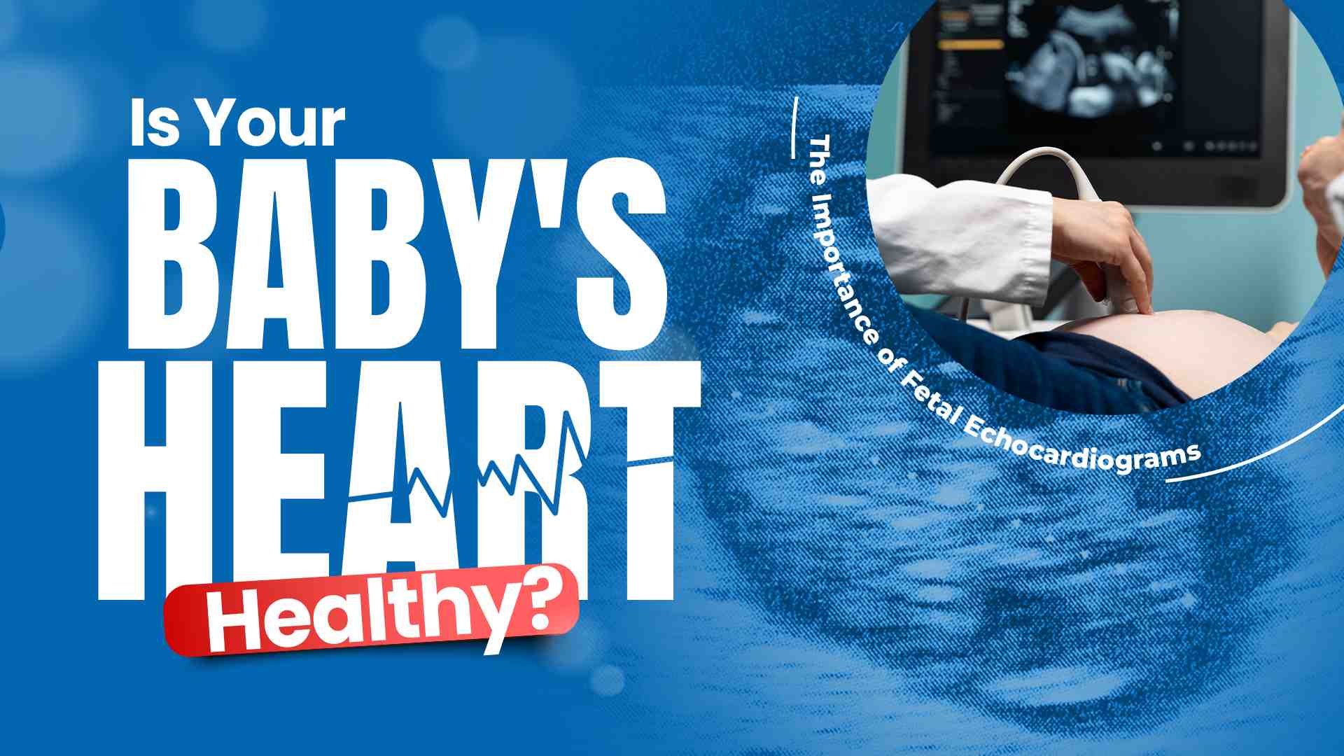
Fetal echocardiogram is ultrasound of baby heart. This thorough scan enables the doctor to assess both atrial and ventricular anatomy and functional characteristics. Similar to a general ultrasound, which delivers rough timings at the fetal organ, anatomical echocardiogram targets most explicitly in and around heart with its galleries of chambers, valves & vessels.
Fetal echocardiogram is ultrasound of baby heart. This thorough scan enables the doctor to assess both atrial and ventricular anatomy and functional characteristics. Similar to a general ultrasound, which delivers rough timings at the fetal organ, anatomical echocardiogram targets most explicitly in and around heart with its galleries of chambers, valves & vessels.
-
Family History of Heart Defects
Any first degree relative of the fetus like parents, sibling or prior pregnancy have history of cardiac anomaly.
-
Abnormal Ultrasound Findings
If your baby's heart appears to be abnormal in routine anomaly scan or there are risk factors for a possible cardiac condition like increased nuchal translucency in first trimester or any extra cardiac abnormality, you will have to undergo detailed fetal echocardiography. This test gives a more detailed image, which helps confirm whether you have heart defect or not.
-
Maternal Causes
If mother has pregestational diabetes or autoimmune disorder like lupus, fetal echocardiography is recommended. During pregnancy, some infections (such as rubella or cytomegalovirus) can have an impact on the formation of a baby's heart. IVF (In vitro fertilisation) pregnancy and any exposure to selected teratogen like carbamazepine etc.
It is performed through a female abdomen with the use of ultrasound like any other regular ultrasound. It is done most often between 18 and 24 weeks of pregnancy although some cardiac structures are better visualised at around 22-24 weeks. The actual test takes from 30 minutes until one hour to complete depending on the position of the baby and if there is any abnormality noted in the scan.
The ultrasound images of the baby's heart are displayed on a monitor as the probe is placed on your abdomen during this procedure.
During the third trimester, several technical limitations can significantly affect the ability to evaluate fetal cardiac anatomy through ultrasound. Maternal obesity can create challenges in sound wave penetration, leading to suboptimal imaging. Additionally, the position of the fetus can obscure views of the heart, especially if the fetal spine or limbs are blocking the line of sight.
Maternal Fetal Medicine Specialist analyzes these images to look for the heart structure and function.
The results of a fetal echocardiogram are usually available immediately after the procedure. Dr. Dash says, " if echo is otherwise normal with demonstration of baby having a good heart development then routine obstetric follow up is needed” If irregularities are found, your healthcare provider will inform you and discuss the results as well as what happens next with their parents.
The test can help diagnose a number of different types of heart defects. Few are enlisted below:
- Situs abnormalities like dextrocardia, heterotaxy.
- Atrio ventricular septal defects
- Tetralogy of Fallot
- Transposition of great arteries
- Ebstein anomaly
- Coarctation of the aorta
- Ventricular septal defects (VSD)
- Hypoplastic left heart syndrome
- Aortic stenosis, pulmonary stenosis
- Complete and congenitally corrected transposition of great arteries
- Common arterial trunk
- Fetal arrythmias
Early detection of heart defects through fetal echocardiograms is crucial for several reasons:
- Treatment planning: Early detection of heart disease makes it easier for health care providers to plan the best course of treatment which may involve surgery, medication or monitoring.
- Parental preparation: If you find out about your baby's heart condition before birth, prepared to learn and get support for a well-informed future.
- Improved outcomes: The earlier children with a congenital heart defect are identified, the better quality of life and long-term survival is likely to be.
A fetal echocardiogram is a powerful diagnostic test that can check the health of your baby's heart during pregnancy. When potential heart problems are discovered in this type of early screening test for your baby, you can be prepared down the line which will influence so much in terms of your babies future and how to go about taking care of him as a whole. If you worry about your baby's heart, talk with your gynaecologist about a fetal echocardiogram.
Kailash Hospital, largest and best Heart Centre in Noida has latest technological support for Fetal Echocardiography. We offer the services of an experienced team of maternal fetal medicine specialists, Dr. Pratima Dash and Dr. Neha Gupta who are committed to providing comprehensive care.


 +91-9711918451
 international.marketing@kailashhealthcare.com


